Untersuchungen von Skelettabschnitten
Rib images (thoracic cage, hemithorax)
Preparation: none
The images are prepared in two planes. Recent or older fractures can be assessed (displacement of fragments in relative to each other, serial rib fractures).
Pathological processes of the ribs, such as tumours or metastases will also be visible (pathological fracture).
Entire spine (cervical, thoracic, lumbar spine)
Preparation: none
The images are done in two planes. They are used primarily to assess intervertebral disc generation, bone spur formation, and slipped vertebrae, as well as transitional vertebrae formations.
Functional images are taken during strong forward bending (anteflexion) and bending backward (retroflexion). These special images can be made of the cervical and lumbar spine.
Pelvis overview image
Preparation: none
Used for assessment of potential pelvis asymmetry, and to examine the hip joints, symphysis, and sacroiliac joints.
Skull x-ray
Preparation: none
Prepared in two planes and used primarily to diagnose injuries to the bone. However, pathological calcification and bone changes (plasmacytoma) can also be detected.
Haemorrhage without bone injury cannot be identified.
Paranasal sinuses
Preparation: none
Three images are made of the paranasal sinuses. Infections and air-fluid levels can be recognized, as well as mucous membrane polyps.
Check-up images following therapy and for possible puncture for air-fluid level.
Long Bones
Preparation: none
- Upper arm
- Forearm
- Thigh
- Lower leg
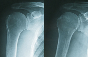
Joints
Preparation: none
- Wrist
- Elbow joint
- Should joint
- Hip joint
- Knee joint
- Ankle
Hand and foot x-ray
Preparation: none
Assessment of malposition, arthrosis, fractures, and bone changes.